[The use of virtual reality (and presence) recently gave a team of doctors the insights they needed to save the lives of conjoined twins; the story is from The Washington Post, where it includes more images and a 3:12 minute video, and more details are available from the University of Minnesota. –Matthew]
How doctors used virtual reality to save the lives of conjoined twin sisters
By Peter Holley
July 23, 2017
During an 18-year career in medicine, Daniel Saltzman — the chief of pediatric surgery at University of Minnesota Masonic Children’s Hospital — has grown accustomed to looking at X-rays as if they were imperfect road maps of the human body.
He compares this exercise to looking at a one-dimensional traffic map on your smartphone and thinking about that image in three-dimensional terms in your mind.
Like any other road map, an X-ray image is an incomplete reduction of reality that can misrepresent challenges or include distortions, which explains why, even in 2017, high-stake surgeries can involve a shocking degree of guesswork and improvisation.
“That’s why medicine is still an art as much as a science,” Saltzman told The Washington Post.
For decades, increasingly sophisticated imaging techniques have allowed doctors to peer into the human body before they cut it open, reducing uncertainty and helping them prepare for complicated procedures. Now, advances in virtual reality may flip that dynamic on its head, allowing doctors to confront the unknown before they even enter the body.
The latest evidence of this revolutionary shift in health care is the successful separation of two conjoined newborn sisters in Minnesota. Until their separation in May, Paisleigh and Paislyn Martinez were attached from their lower chest to their bellybuttons — a condition known as thoraco-omphalopagus. Both babies survived the dangerous, nine-hour procedure, a development that Saltzman and other surgeons involved link directly to their use of virtual reality before surgery.
“It felt like I was working in the future,” Saltzman said. “It was extraordinarily exhilarating.”
Conjoined twins are extremely rare, occurring as infrequently as one of 200,000 live births to up to one in a million births, depending on how the babies are attached, according to University of Minnesota doctors.
Separating them is a risky procedure, though survival rates differ depending on how the siblings are connected and which organs they share, according to the University of Maryland Medical Center. The Medical Center notes that twins “joined at the sacrum at the base of the spine have a 68 percent chance of successful separation, whereas, in cases of twins with conjoined hearts at the ventricular (pumping chamber) level, there are no known survivors.” In the latter case, the hearts are completely joined.
Experts said they were unaware of any other example of virtual reality being used to prepare for the separation of twins partially conjoined at the heart. Virtual reality has been used to assist in the separation of twins conjoined at the head on three occasions, two of which were carried out by Housing and Urban Development Secretary Ben Carson, experts said.
Anthony Azakie — chief of pediatric cardiac surgery and co-director of the Heart Center at the University of Minnesota Masonic Children’s Hospital — called the university’s procedure a “once-in-a-lifetime event.”
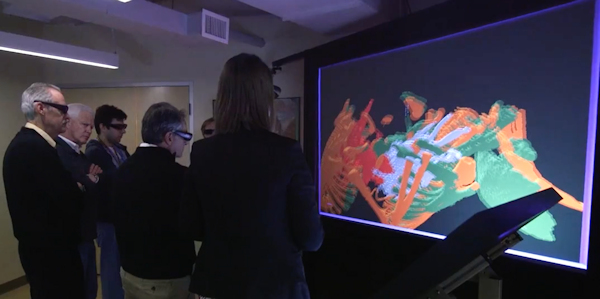
Using goggle-like virtual reality glasses a month before surgery, Saltzman, Azakie and their team were able to explore a 3-D model of the twins’ hearts, virtually embedding themselves inside the walnut-sized organs as if the infant’s anatomy had been blown up to the size of a living room.
“It’s completely surreal and the resolution is unbelievable,” Azakie said. “The details are absolutely superb.”
The experience was not only riveting, but revelatory, doctors said, so much so that the stunned surgeons decided to alter their entire operative strategy. Within minutes after putting on the glasses, Saltzman and Azakie discovered something unexpected: new connective tissue — a “bridge” — linking the girls’ intertwined hearts, one of which had become heavily reliant upon the other to filter impurities and remain beating because of a severe congenital heart defect.
That defect meant that the lives of both babies were in jeopardy and doctors would have to conduct the surgery several months early, before the twins were as robust and healthy as doctors had hoped they’d be.
Standing inside the 3-D rendering of the infants’ hearts, the challenge before the physicians was daunting. They realized that improperly severing that connection could led to the twins bleeding to death. Placing pressure on the hearts could cause blood loss or arrhythmia so severe the organs stopped beating entirely. They needed to find a way to navigate around the connection that didn’t damage each delicate organ.
The team members interacted with the 3-D model using a “track system” that allowed them to turn their heads without distortion. Doctors said the ease of the virtual interaction helped the team arrive at a simple, yet elegant solution, which they drew up on a whiteboard moments later. The surgeons decided to flip the babies around on the operating table so that the procedure occurred from the opposite angle. In the end, doctors said, the straightforward solution to a complicated quandary may have saved the twins’ lives.
“In our line of work — especially in pediatric cardiac surgery — it’s important that one is able to think on their feet and plan for the unexpected,” Azakie said. “The imaging helped us prepare by developing an approach in the event that we came across something we didn’t expect.”
“It was like the Oculus Rift of pediatric heart surgery.”
The planning effort to get to that point was methodical, with weekly meetings, endless tests and trial runs with a team of nearly 50 hospital staffers who practiced each stage of the surgery using baby dolls that had been sewn together.
To create the virtual model of the infants’ hearts, Saltzman and Azakie and other team members partnered with the University of Minnesota’s Earl E. Bakken Medical Devices Center, where experts used software to turn MRI’s and CT scans from both infants into a detailed virtual model. The university’s Visible Heart Lab also created a 3-D printed model of the hearts using a printer purchased online for $300.
Bethany Tourek, a PhD candidate in the University of Minnesota’s department of mechanical engineering, said watching the medical team experience the virtual heart for the first time was a seminal moment.
“My favorite part was actually watching these four medical doctors standing inside this screen and being in complete silence at first,” Tourek said. “You could just see them thinking and processing and taking it all in for the first time. It was a wonderful experience.”
Two months after their separation, the twins are still recovering, but doctors say that they’ll lead healthy, independent lives, with a scar on their chests the only evidence that they were ever conjoined in the future.
Doctors said they expect the 3-D modeling and imaging used to see the twins’ anatomy to appear in medical journals, setting a precedent for surgeries that ripples beyond procedures involving conjoined twins.
“Separating these infants was no small feat,” Saltzman said. “The fact that we got to do it using virtual reality for direct patient care makes that feat truly incredible.”